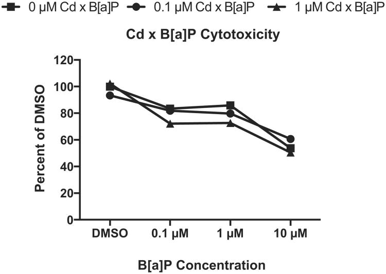
Fig. 5.
Co-exposure to Cd and B[a]P resulted in a significant increase in cytotoxicity at the highest dose only. After 24 h of either 0 μM Cd, 0.1 μM Cd, or 1 μM Cd, cells were exposed to concentrations of B[a]P for 24 h. Only groups exposed to 1 μM Cd × 10 μM B[a]P significantly differed from untreated controls, p < 0.001. Viabilities are expressed as percent DMSO control as determined by the MTT viability assay. Points represent mean ± SEM (n = 8).

