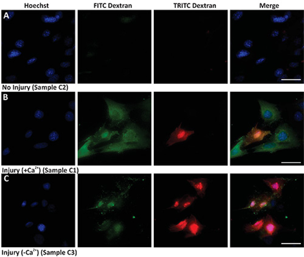
Figure 1. Bulk cell membrane injury by glass beads.
Images show nuclear staining (Hoechst, left panel), injury (green staining - FITC Dextran), unrepaired cells (red staining - TRITC Dextran), and merged images (Merge). (A) Uninjured cells in the presence of calcium, (B) Injured cells in presence of calcium, and (C) Injured cells in absence of calcium. Scale bar 10 µm.

