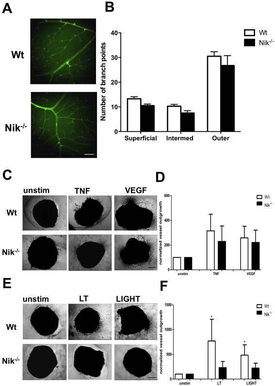
Figure 4. Genetic deletion of NIK results in reduced pathological angiogenic responses.
A, Representative retinas from Wt and Nik−/ mice, stained with isolectin B4 (green) (scale bar for both panels, 100 μm). B, Quantification of vessel branching in the superficial, intermediate and outer vascular plexus. C, Aortic rings of Wt and Nik−/− mice cultured in medium alone, TNF-α or VEGF. Representative pictures are shown (scale bar all panels, 500 μm). D, Quantification of TNF-α and VEGF-induced aortic vessel outgrowth. E, Representative pictures of unstimulated, LT- or LIGHT-stimulated aortic rings (scale bar all panels, 500 μm). F, Quantification of LT- and LIGHT-induced aortic vessel outgrowth. Panels B,C,D n=4 per group, 2 independent experiments. Panels A,E,F n=5 per group, 2 independent experiments. Panels B,D,F, represent mean±SEM *<P0.05.

