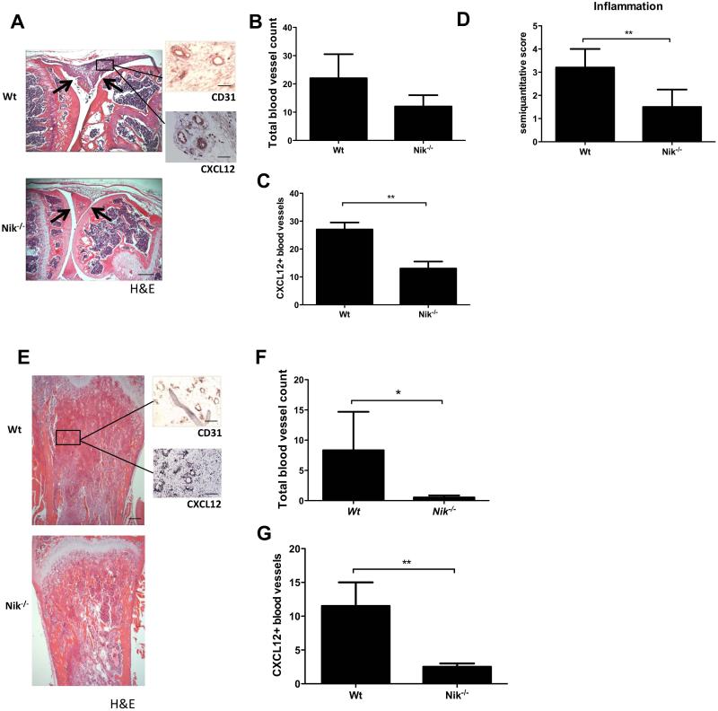
Figure 5. Nik−/− mice exhibit reduced inflammation-induced angiogenesis in arthritis and reduced tumor-associated angiogenesis in a melanoma model.
A, Representative picture of a mBSA-injected Wt mouse knee joint showing a severe inflammatory infiltrate (between arrows), in contrast to a mBSA-injected Nik−/− knee joint showing a reduced inflammatory infiltrate and intact cartilage (between arrows; scale bar 500 μm). Sagittal sections of knee joint with H&E staining and CD31 or CXCL12 IHC staining (insets; scale bars 100 μm). B, Quantification of the total number of synovial blood vessels in Wt and Nik−/− mice. C, Quantification of CXCL12+ blood vessels in synovial tissue of Wt and Nik−/− mice. D, Quantification of synovial inflammation on a semiquantitative scale of 0 to 4. E, Representative pictures of tibial sections from Wt and Nik−/− mice, with B16 melanoma tumor tissue in trabecular bone. H&E-stained sections of tibias show marrow replacement by tumor cells beneath the growth plate (scale bar 500 μm), and insets show CD31 and CXCL12 IHC staining on tumor tissue (scale bar insets, 100 μm). F, Quantification of the total number of blood vessels in melanoma tumor tissue in Wt and Nik−/− mice. G, Quantification of CXCL12+ blood vessels in melanoma tumor tissue of Wt and Nik−/− mice. Panels A-D: n=5 per group; Panels E-G: n=6 per group. Panels B-D,F,G represent mean±SEM *<P0.05, **P<0.001.

