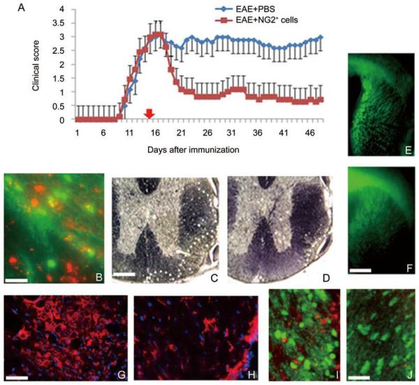
Fig. 2.
Purified NG2+ cells ameliorate EAE, migrate into the demyelinated CNS, promote remyelination and reduce inflammatory T cell infiltration. A: Injection of 1×10⁶ NG2+ cells into mice with MOG35-55-induced EAE resulted in significant neurological improvement compared to control animals that received Pbs. b: Labeled NG2+ cells (red) were found in lesion areas of white matter identified by reduced expression of PLP-EGFP 24 h after injection. c and D: the lesion load was reduced in the spinal cord of animals that received NG2+ cells compared to controls as shown by black gold staining. the relative area of demyelination at the periphery of the spinal cord is delineated by white dots. E and F: treatment with NG2+ cells promoted endogenous myelination in PLP-EGFP host animals with EAE. Control animals with EAE (E) showed significantly reduced EGFP expression in the corpus callosum and forebrain compared to animals treated with NG2+ cells (F). G and H: Demyelinated lesion areas in the spinal cord of control animals (G) were characterized by infiltrating CD3+ t cells that were substantially reduced in animals treated with NG2+ cells (H). I and J: The number of endogenous oligodendrocytes (green) increased in regions of demyelination that contained NG2+ cells (red) (I) compared to regions devoid of NG2+ cells (J). Scale bars: B, 50 μm; C–F, 500 μm; G–J, 100 μm.

