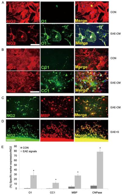
Fig. 3.
Adult NG2+ cells generate mature oligodendrocytes in response to cues from demyelinating tissue. Adult spinal cord NG2+ cells grown in the presence of EAE-conditioned medium for 3–5 days generated increased numbers of differentiated mature oligodendrocytes that were O1+ (A), CC1+ (B), and MBP+ (C) compared with parallel cultures grown in control medium. When 1 × 10⁴ NG2+ cells were co-cultured with 1-mm slices from EAE brain tissue (EAE-S) for 3 days, higher numbers of CNPase+ cells differentiated from the NG2+ cells (D). Quantification of cells differentiated from the NG2+ cells in the presence of EAE-cM or EAE-s is shown in E. *Control versus EAE-cM/s: O1 P <0.05, CC1 P <0.05, MBP P <0.05, CNPase P <0.001. Mean ± SEM of duplicate preparations from three independent experiments. Scale bars in A–C, 150 μm and D, 300 μm.

