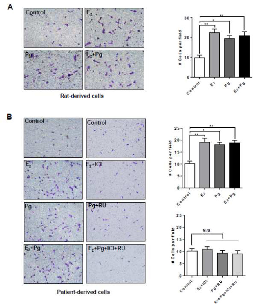
Figure 2. Progesterone increases the migration of TSC2-deficient cells.
(A) Rat-derived cells and (B) Patient-derived cells were treated with 10 nM E2, 10 nM progesterone (Pg), 10 nM E2 + 10 nM Pg, or vehicle control for 24 hr. 20,000 cells were seeded in the upper chamber in the presence of steroids or vehicle. Cell migration was measured using the Boyden chamber transwell assay. Number of cells migrated through the chamber after 24 hr incubation was detected by Crystal Violet staining and quantitated. (B) Patient-derived cells were pre-treated with ICI 182,780 (ICI, 10 µM) or RU-486 (RU, 20 µM) for 12 hr, and then stimulated with 10 nM E2, 10 nM Pg, 10 nM E2 + 10 nM Pg, or vehicle control for 24 hr. Cell migration was measured using the Boyden chamber transwell assay. Number of cells migrated through the chamber was detected by Crystal Violet staining and quantitated. Data are mean ± SEM, n = 3 in triplicate. * p < 0.05, ** p < 0.01, Student t test.

