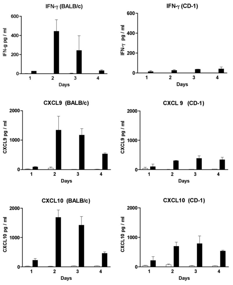
Fig. 4.

Identification of IFN-γ, CXCL9 and CXCL10 in the sera of MHV infected mice by EIA. Sera from 3 to 4 mice were tested at each time point (day 1,2,3 and 4). MHV infected mice are represented by the black bars while mock injected mice are represented by the white bars.
