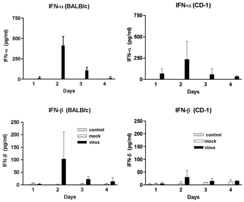
Fig. 6.

Identification of IFN-α and IFN-β in the sera of MHV infected mice by EIA. Sera from 3 mice were tested at each time point. MHV infected mice are represented by the black bars, mock injected mice are represented by the white bars and control mice are represented by the hashed bars.
