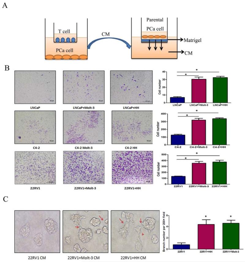
Figure 3. Co-culture of T cells promoted PCa invasion.
A.CMs collected from PCa/T cells co-culture could promote PCa invasiveness using in vitro matrigel invasion assay. The PCa/T cells were co-cultured in 0.4 μM transwell plate for 2 days. The CM or control media were collected, diluted 1:1 with 10% FBS RPMI media, and placed into the lower chamber of transwells. The 1×10⁵ PCa cells (without any pre-treatment) were plated onto upper insert chamber pre-coated with matrigel for 36 hr invasion assay. B. Toluidine blue staining results showed the PCa/T cells co-cultured CM can promote PCa cell invasion. Invasion assay has been performed on 3 different PCa cell lines, LNCaP, C4-2 and CWR22RV1(22RV1) and 2 different T cells, Molt-3 and HH. (*P<0.05).C. 3D invasion assay showed that more acini-like structures formed when treating the parental CWR22RV1 cells with CWR22RV1/T cell co-culture CM (200×) (*P<0.05).

