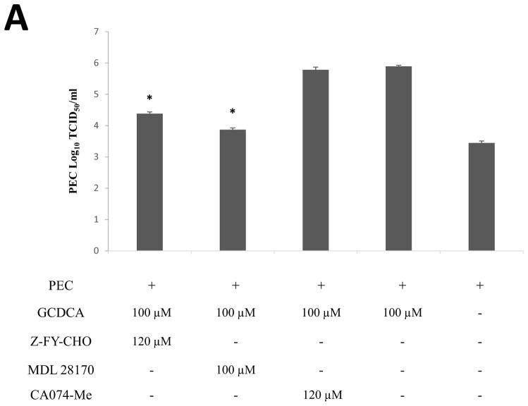
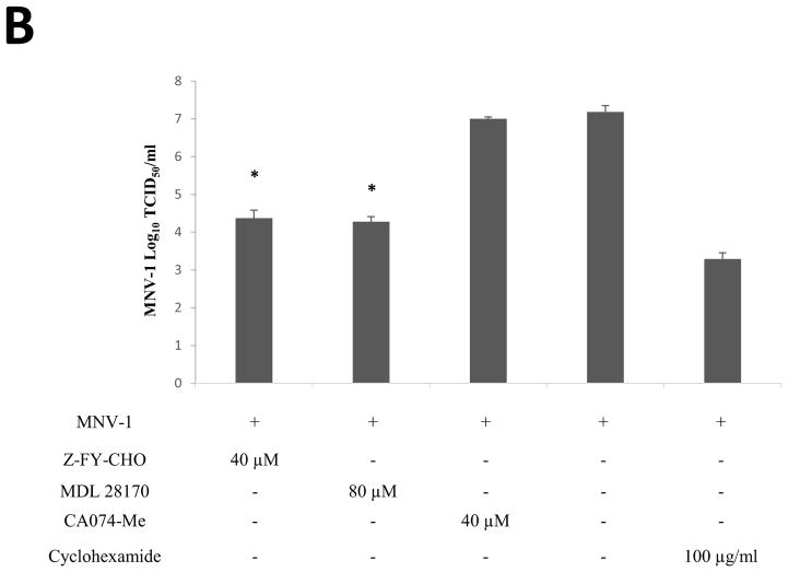
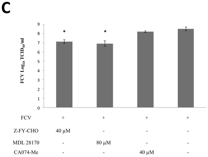
Figure 1. Effects of cathepsin inhibitors in the replication of PEC, MNV-1 or FCV.
LLC-PK, RAW267.4 or CRFK cells were incubated with cathepsin L inhibitors, Z-FY-CHO and MDL 28170, or a cathepsin B inhibitor, CA074-Me, for 1h, then infected with (A) PEC (MOI 50) (B) MNV-1 (MOI 10) or (C) FCV (MOI 50) in the presence of each inhibitor. For PEC, GCDCA (100 μM) was present in the media to support virus replication during virus infection. Following virus infection for 1 h, cells were washed and the media was replaced with fresh media containing an inhibitor. Cells were further incubated at 37°C and collected at 12 h PI. Viral replication was quantified by TCID50 assay. Asterisk indicates that virus titer was significantly reduced by an inhibitor compared to the control (P < 0.05).


