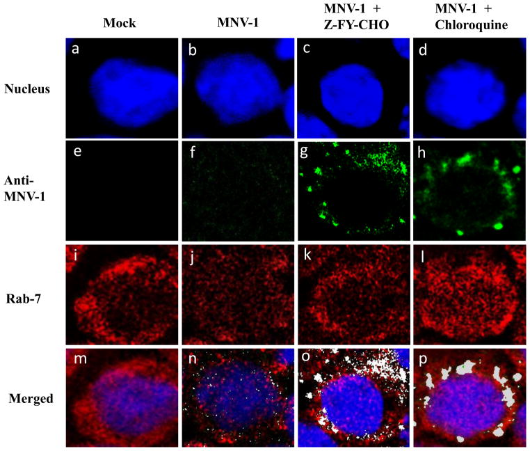
Figure 6. The effects of cathepsin L inhibitor or chloroquine in MNV-1 entry into the cells.
RAW267.4 cells were pre-treated with mock (medium), Z-FY-CHO or chloroquine for 30 min before MNV-1 inoculation (MOI 10). Following virus infection at 37 °C for 1 h, cells were fixed and probed with rabbit polyclonal anti-Rab7 or guinea pig polyclonal antibody against MNV-1, followed by PerCP-Cy5.5 labelled secondary antibody against Rab7 (red, i to l) or FITC-labelled secondary antibody against MNV-1 (green, e to h). Nuclei were stained with sytox orange (pseudo colored blue, a to d). Confocal images on the prepared samples were obtained and colocalization analysis of Rab7 and MNV-1 was performed by ImageJ software. In the merged images (p to t), colocalization of MNV-1 (green) and Rab7 (red) appears in white color.

