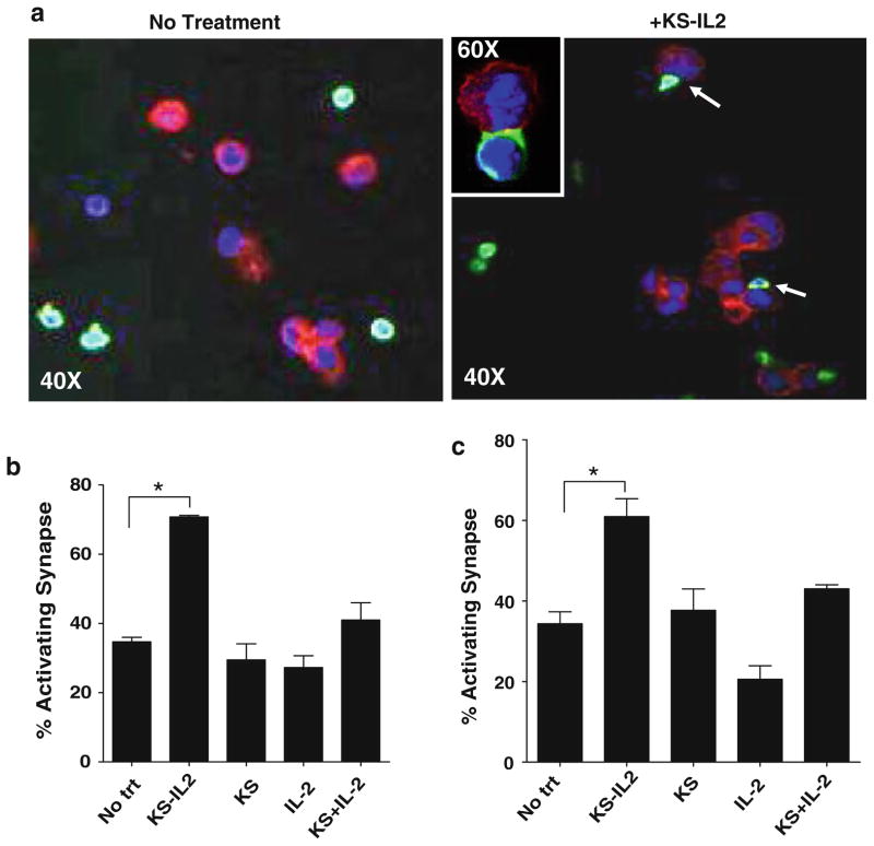
Fig. 1.
huKS-IL2 increases AIS between primary NK cells and ovarian tumor cells. a Naïve NK cells from peripheral blood of healthy donors were incubated with OVCAR-3 cells in the presence of IC (+huKS-IL2) or absence (no treatment). Cells were fixed onto coverslips and stained for AIS markers LFA-1, CD2 and F-actin. Red = F-actin, Blue = nucleus, Green = LFA-1. OVCAR-3 cells are seen stained mainly as cells with red cytoplasm, while the smaller NK cells are identified by their LFA-1 (green) expression. When huKS-IL2 is present (Right photomicrographs in a “+huKS-IL2”) several NK cells are seen forming synapses with the OVCAR-3 cells. Increased density of LFA-1 is seen at sites of AIS formation (white arrows). Inset in a demonstrates a higher power (60× + zoom) view of an AIS. Pictures were taken using a confocal microscope at 40×. Pictures are representative of four separate donors. AIS formation was quantified by counting NK-OVCAR-3 conjugates showing polarization of LFA-1 (b) or CD2 (c). AIS formation was determined when NK cells and OVCAR-3 cells were incubated in media (no treatment) or media containing huKS-IL2, huKS, IL-2, or huKS + IL2. Percentage of activating synapses was determined as described in Methods. Quantification of coverslips by fluorescent microscopy, shown in the bar graphs, indicates increased AIS when huKS-IL2 is added. Bar graphs are mean of data obtained using NK cells from four individual healthy donors. *P <0.05

