Fig. 2.
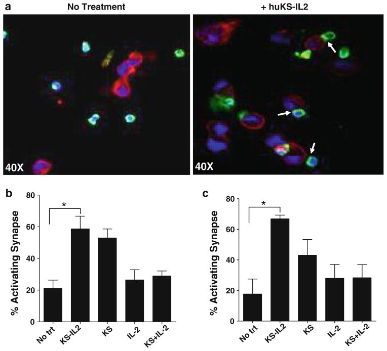
Enhanced IC-mediated AIS formation between targets and naïve NK cells from peripheral blood of ovarian cancer patients. Naïve NK cells were isolated from peripheral blood of four ovarian cancer patients at the time of diagnosis of the disease. All experiments were conducted similar to those with NK cells from healthy donors (Fig. 1). Photomicrographs (a) show increased AIS formation in the presence of huKS-IL2. Polarization of LFA-1 (b) and CD2 (c) were used as indicators of AIS formation. All other details are same as those provided in the legend for Fig. 1. *P <0.05

