Fig. 7.
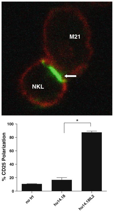
CD25 polarizes at the immune synapse. a M21 (melanoma) cells and NKL cells were incubated together with either no treatment, hu14.18, or hu14.18-IL2 for 25 min. Cells were fixed onto a glass coverslip, stained for CD25 (green) using a non-blocking antibody. Cells were visualized using confocal microscopy. Arrow indicates polarization of CD25 between NKL and M21 cells. Images were taken at 40× and digitally zoomed. b Polarization of CD25 was quantified by dividing the number of conjugates between NKLs and M21 s where CD25 was polarized at the synapse by the total number of conjugates. *P < 0.02

