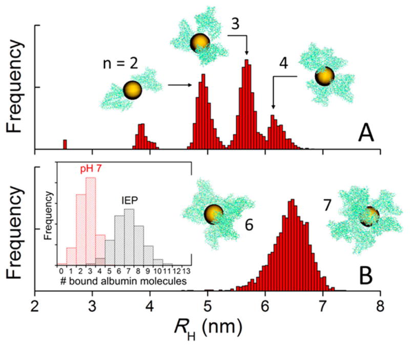
Figure 6.

Hydrodynamic radii (RH) and morphology of AuNP/albumin complexes at physiological pH (A) and at the isoelectric point (IEP) (B) in a diluted nanoparticle solution at 37 °C and physiological albumin concentration, obtained from canonical Monte Carlo simulations. The nanoparticles have a diameter of 5 nm. The geometric arrangement of proteins and the number (n) of proteins bound to the nanoparticle (histograms in inset of panel B) depend on the charge distribution on the protein, which is controlled by the pH and the ionic strength of the solution. The aggregates shown in the insets are snapshots taken from the equilibrated simulations and are drawn to scale (yellow, AuNPs; green, atomic representation of albumin obtained after fine graining).
