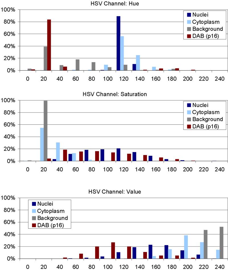
Figure 3. Analysis of objects in HSV color space.
340 images of nuclei, cytoplasm, background, and p16 stained cells (85 images each) have been analyzed for their composition in the HSV color model after conversion from the original Red-Green-Blue (RGB) images. From each image 10 pixel have been collected, yielding a training data set of 3400 pixels from which the classifiers for nuclei, cytoplasm, and p16 stain were developed. In each channel (Hue, Saturation, Value) distinct distributions can be identified allowing discriminating of the image sets.

