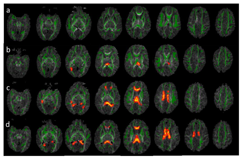
Figure 3.

DTI TBSS results show that the more previous brain hemorrhage, the more white matter microstructural abnormalities (as reflected by more regions with lower FA compared to controls). Green represents the average white matter skeleton overlaid on FA images, orange/yellow represent white matter regions with significantlylower FA values (when compared to control infants, p<0.05, fully corrected for multiple comparisons) in a) ELBW infants with no old blood product deposition on MRI at term-equivalent age, b) ELBW infants with old blood product deposition on MRI but no ultrasound IVH diagnosis after birth, c) ELBW infants with old blood product deposition on MRI and previous ultrasound diagnosis of grades 1 or 2 IVH, and d) ELBW infants with old blood product deposition on MRI and previous ultrasound diagnosis of grades 3 or 4 IVH.
