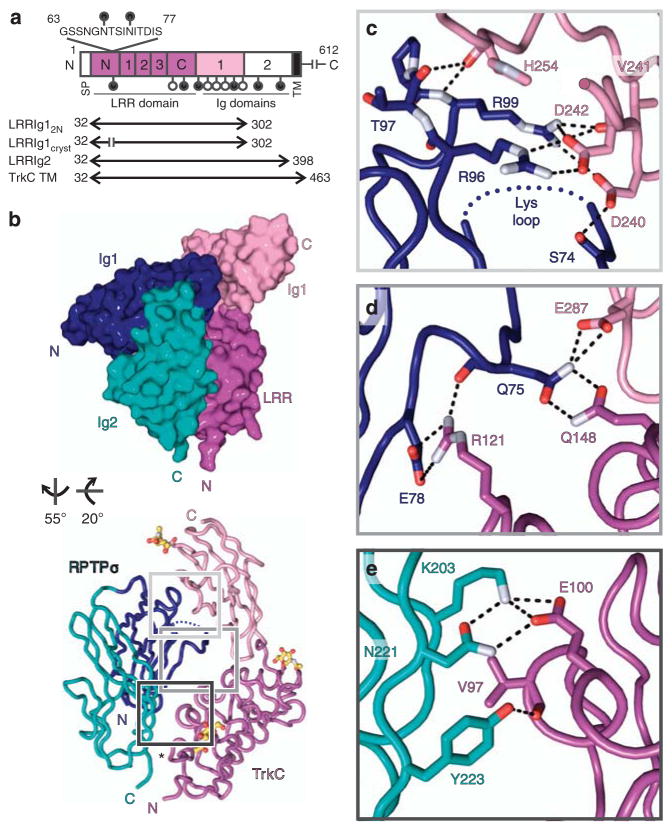
Figure 2. Trans-synaptic RPTPσ:TrkC complex crystal structure.
(a) TrkCTK- (non-catalytic isoform) domain organization. LRR, leucine-rich repeat region (N, N-terminal cysteine-rich region; 1–3, leucine-rich repeats; C, C-terminal cysteine-rich region). Putative N-linked glycosylation sites, lollipops; filled lollipops remain in LRRIg1cryst construct. (b) Space-filled and tube representations of chicken RPTPσ Ig1–2:TrkC LRRIg1cryst crystal structure. N-linked glycans in atom representation. Disordered RPTPσ Lys-loop, blue dotted line; TrkC LRRIg1cryst amino-acid residue 62–78 junction, asterisk. (c–e) Detailed view of bonding interactions at RPTPσ:TrkC interface for binding sites 1–3. Corresponding electron density is illustrated in Supplementary Fig. 7. Potential electrostatic and hydrogen bonds, black dashed lines; oxygen atoms, red; nitrogen atoms, bluewhite.

