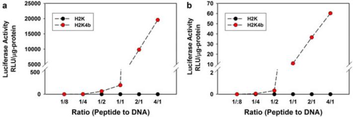
Figure 2.
in vitro transfection. After MDA-MB-435 cells reached 70% confluency in 24-well plates, increasing amounts (0.125, 0.25, 0.5, 1.0, 2.0 and 8.0 μg) of peptides (H2K or H2K4b) were mixed with 1.0 μg of the plasmid (CPG-Luc) in Opti-MEM (a) or water (b) and the mixture was allowed to stand for 30 min. After the transfection complex was added to cells for 24 h, the cells were lysed and luciferase activity was measured. Duplicate measurements were made at each concentration in four separate experiments. Data from the luciferase activity is expressed as the mean. A 1/1, w/w ratio is approximately equivalent to a 1.3/1, N/P ratio.

