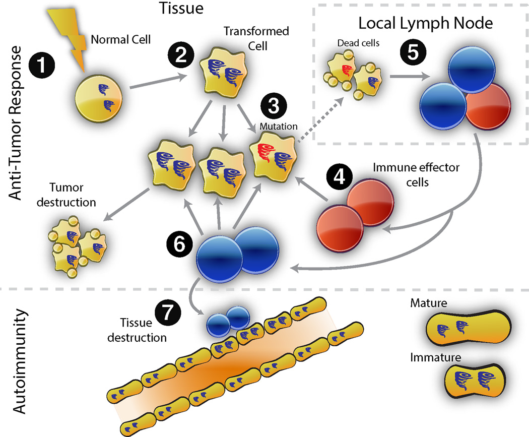
Figure 1. Model for cancer-induced autoimmunity.
Transformation of normal cells (1) may result in gene expression patterns which resemble immature cells involved in tissue healing (2). Occasionally, autoantigens become mutated (3); these are not driver mutations, and not all cancer cells have them. The first immune response is directed against the mutated form of the antigen (4), and may spread to the wild type version (5). Immune effector cells directed against the mutant (depicted in red) delete exclusively cancer cells containing the mutation (6). Immune effector cells directed against the wild type (blue cells) delete cancer cells without the mutation, and also cross-react with the patient’s own tissues (particularly immature cells expressing high levels of antigen, found in damaged/repairing tissue). Once autoimmunity has been initiated, the disease is self-propagating. Immature cells (expressing high antigen levels) that repair the immune-mediated injury themselves become the targets of the immune response, sustaining an ongoing cycle of damage/repair that provides the antigen source fueling the autoimmune response.

