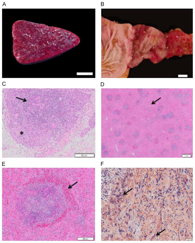
Fig. 3.
Gross and microscopic lesions of SHF. (A) Spleen, cut surface (JM-24054). White pulp is markedly diminished within diffusely expanded red pulp, bar=1 cm. (B) Duodenum (JM-22015). The mucosa exhibits ecchymotic to diffuse hemorrhage that extends to the pyloric valve, bar=1 cm. (C) Thymus stained with H&E (JM-24054). There is diffuse lymphocyte necrosis of cortical lymphocytes (asterisk) and depletion of medullary lymphocytes (arrow), bar=200 μm. (D) Spleen stained with H&E (JM-24054). Depleted and necrotic follicles are rimmed with a zone of hemorrhage. Red pulp is markedly expanded (arrow), bar=1 mm. (E) Spleen stained with H&E (JM-24054). A splenic follicle exhibiting lymphocyte necrosis within the central portion of the germinal center, the mantle and marginal zones with an adjacent rim of hemorrhage (arrow), bar=200 μm. (F) Spleen (JM- 24054). Splenic red pulp is filled with abundant fibrin strands stained purple by phosphotungstic acid hematoxylin (arrows), bar=20 μm.

