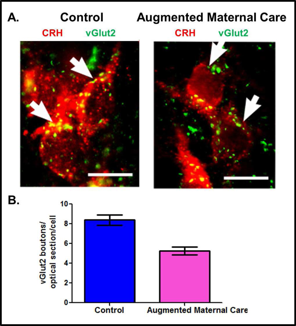
Figure 3. Fewer excitatory (vGlut2-positive) boutons contact CRH-ir neurons in parvocellular PVN of pups that experienced augmented maternal care as compared to the controls.
A. Merged confocal microscope images of sections at the level of the paraventricular nucleus (PVN) of the hypothalamus double labeled for CRH (red) and vGlut2 (green) in P9 rats that had either control or augmented early-life experience. B. Quantification of the vGlut2-positive boutons contacting CRH-ir soma shows a 36% reduction in the vGlut-2 abutting CRH neurons in pups that experienced enhanced maternal care (5.4±0.9) as compared with controls (8.4±0.9). Scale bars, 10µm. Adapted from (Korosi et al., 2010) with permission.

