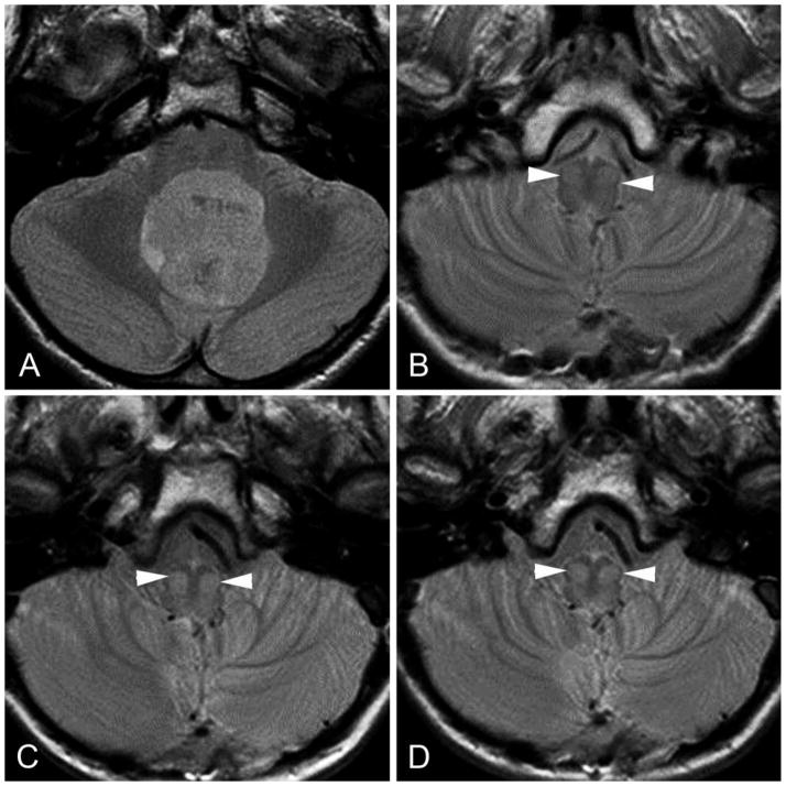
FIG 3.
Transverse proton density–weighted images showing chronological changes (increasing conspicuity) within the bilateral inferior olivary nuclei (white arrowheads) in a patient with PFS during the first year after surgery. A, preoperative study. B, at 1-month follow-up. C, at 6-months follow-up. D, at 10-months follow-up.

