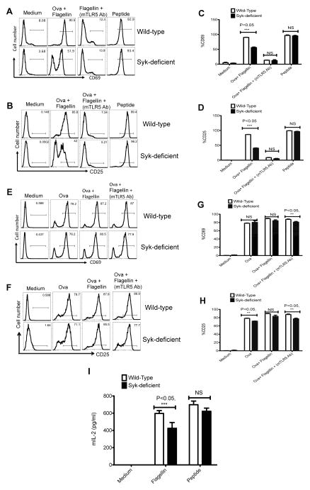
Figure 5. Syk-deficient DCs display a specific deficiency in the activation of flagellin specific CD4 T cells.
CD11c+ DCs (1×10⁵) isolated from wild-type and Syk-deficient chimeric mice were either pretreated with monoclonal anti-TLR5 antibody for 60 min or left untreated and were then cultured with 1×10⁵ SM1 or OT-II T cells for 16 h in the presence of OVA (100 μg/ml), OVA (100 μg) + flagellin (1ng/ml) or flagellin peptide (6μM). A,B) Upper FACS plots (A) and bar graphs (B) show the expression of CD69 after gating on (CD4+CD90.1+) SM1 T cells and lower plots along with bar graphs shows the expression of CD25 (C–D) on gated SM1 T cells. E–H) FACS plots in the upper and lower panel, and bar graphs shows the expression of CD69 (E–G) and CD25 (F–H) on gated OT-II T cells (CD4+CD90.1+) cultured with treated CD11c+ DCs enriched from spleen of wild-type and Syk-deficient chimeric mice as described above. Bar graphs show the percentage of positive cells +/− SEM. 5I) Graph showed IL-2 cytokine measured in the culture supernatant of DC-T cell co-culture experiment (16h) by ELISA. The values represented show mean +/− SEM. Data are representative of three separate experiments done in triplicate. P value <0.05 (***) is statistically significant.

