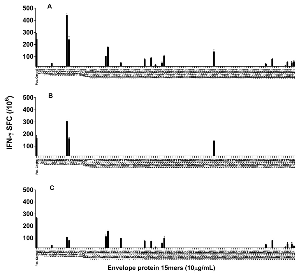
Figure 1.
Response of total splenocytes and CD4+ and CD8+ T cells to the peptides of the YF envelope protein. BALB/c mice were immunized on day 0 and boosted on day 21 with 104 PFUs of the human 17DD YF vaccine, and the splenocytes were tested in IFN-γ ELISPOT assays 7–10 days after the boost. The peptides used for in vitro stimulation were 15mers, overlapping by 11 amino acids and comprising the length of the envelope protein (10 µg/mL). (A) splenocytes; (B) CD4-depleted splenocytes; (C) CD8-depleted splenocytes. Figures represent the average of two to four experiments performed with a pool of three to five mice each. Bars indicate the mean ± SD.

