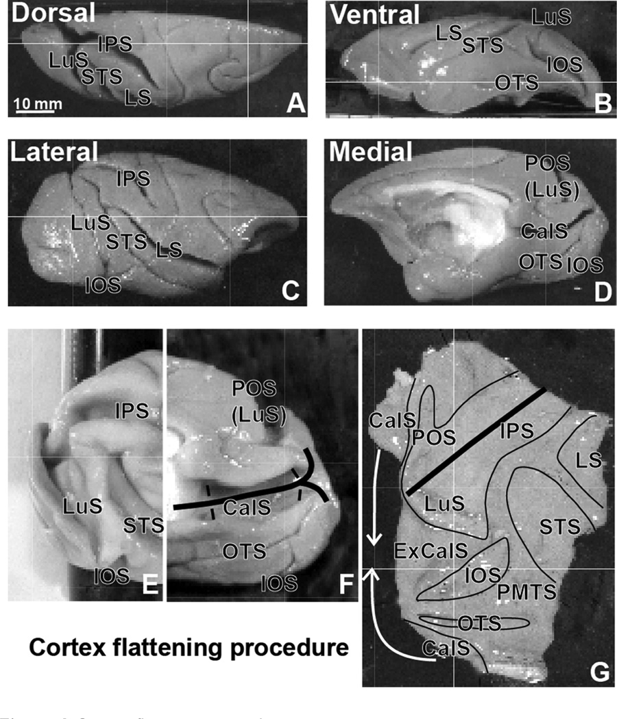
Figure 1. Cortex fattening procedure.
Notes: (A–D) Dorsal, ventral, lateral, and medial views of the right hemisphere with marked sulci. (E–G) The procedure used to fatten the visual cortex of the caudal part of the hemisphere. In (F and G), thick black lines mark cuts along the calcarine sulcus on the medial wall and the IPS on the dorsal aspect of the hemisphere. In (G), thin black lines mark contours of opened sulci. Copyright © 2005. Oxford University Press. Adapted from Stepniewska I, Collins CE, Kaas JH. Reappraisal of DL/V4 boundaries based on connectivity patterns of dorsolateral visual cortex in macaques. Cereb Cortex. 2005;15(6):809–822 by permission of Oxford University Press.21
Abbreviations: CalS, calcarine sulcus; ExCalS, external CalS; IOS, inferior occipital sulcus; IPS, intraparietal sulcus; LS, lateral sulcus; LuS, lunate sulcus; OTS, occipitotemporal sulcus; PMTS, posteromedial temporal sulcus; POS, parieto-occipital sulcus; STS, superior temporal sulcus.

