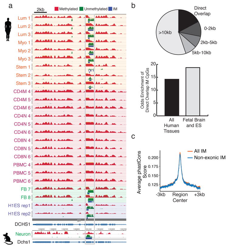
Figure 4. The IM state is conserved in syntenic loci in mouse.
(a) A novel, tissue-specific human IM region in an internal exon of DCHS1 shows conserved IM state at the orthologous exon in mouse. Height for all tracks shows a signal range of 0–50 reads. (b) The pie chart indicates the distance to the nearest human IM region from each aligned mouse IM region. The bar graph shows the fold-enrichment of overlap between human and mouse IM regions at the CpG level using the complete set of human IM regions and a set restricted to cell types analogous to those in the mouse IM analysis. (c) Average phastCons conservation scores over all IM regions and regions that do not overlap coding exons. Scores are based on alignment of 46 vertebrate species.

