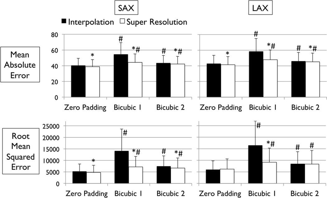
FIGURE 4.
Error measurements vs. high-resolution image. Values are mean ± SD. Black and white bars represent interpolated and super resolution images, respectively. The sample size was n=1,000 for short-axis (SAX) images, and n=500 for long-axis (LAX) images. *: P<0.001 vs. Interpolation; #: P<0.001 vs. zero Padding.

