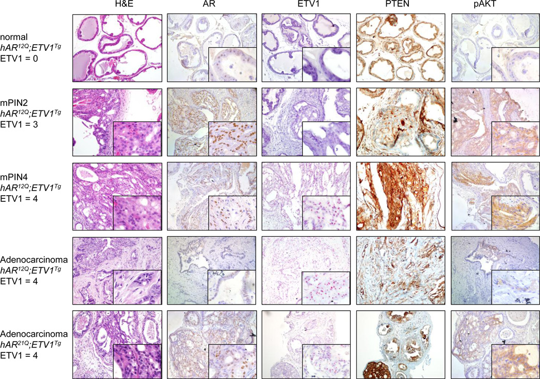
Fig. 5.
Histopathology in Pten-hemizygous mice. Representative prostate sections are shown from, in order, intact hAR12Q;ETV1Tg;Pten+/− mice with normal, PIN2 or PIN4 DLP, as well as macroscopic DLP tumors (adenocarcinoma) from hAR12Q;ETV1Tg;Pten+/− and hAR21Q;ETV1Tg;Pten+/− mice. Sections were stained with H&E. Immunohistochemical (IHC) staining was performed with antibodies to AR, PTEN or pAKT protein (brown staining). In situ hybridization (ISH) was performed with a probe against the human ETV1 transcript (red staining). ETV1 expression level is scored as 0–4, with 4 being highest. Images are shown at 10x-20x magnification with 60x inset. Consecutive sections were used when possible, and each row of images shows stained sections from a single mouse prostate.

