Fig. 9.
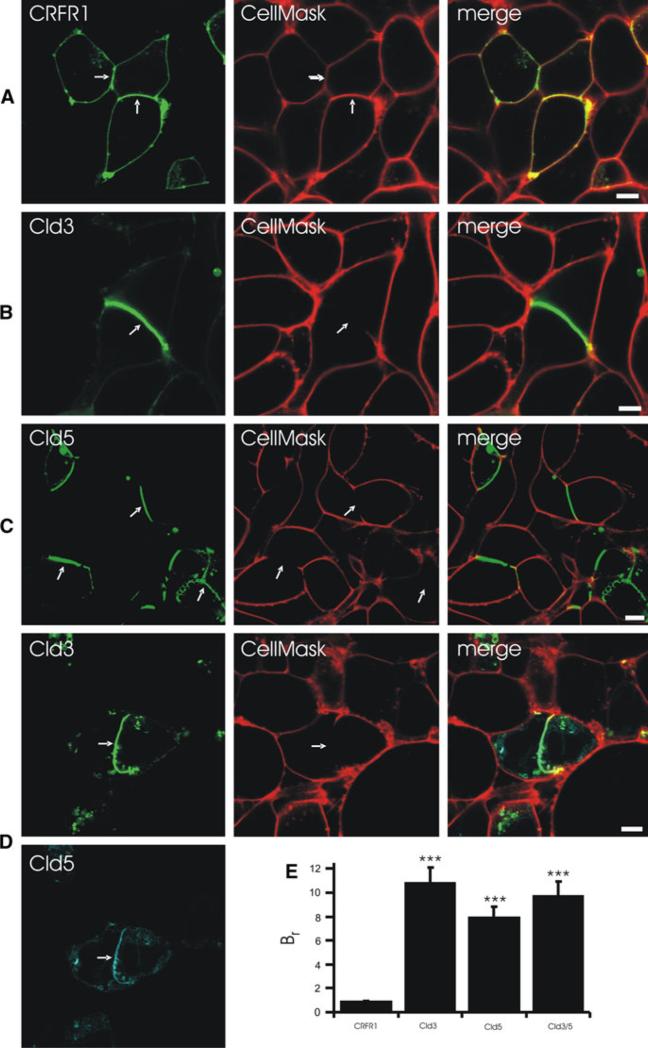
Claudins form diffusion barrier at contacts between claudin-expressing HEK cells. Cells were incubated with CellMask for 5 min and the labeling of the cell membrane was analyzed by confocal microscopy. a CellMask labeled contacts between CRFR1-CFP(control)-expressing and non-expressing cells as well as contacts between two CRFR1-CFP-expressing cells. At contacts between Cld3-YFP- (b), Cld5-YFP (c) or Cld3-YFP/Cld5-CFP- (d) expressing cells, CellMask labeling was much weaker or absent. e The barrier ratio (Br), as a measure for paracellular barrier preventing CellMask labeling, was determined using confocal intensity profiles. Bar, 5 μm; ***p < 0.001 versus CRFR1; arrows, contacts between cells expressing fusion proteins

