
Figure 2. Age-related changes in transverse temporal white matter regions.
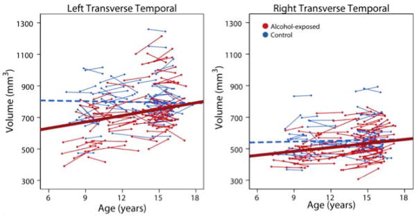
The only areas with significant age-group interactions were the bilateral transverse temporal regions, in which alcohol-exposed subjects (red) showed significant volume increases over time, but no significant changes were noted in control subjects (blue). The best fit line for control subjects (dotted blue line) was not significant and is just shown for reference.