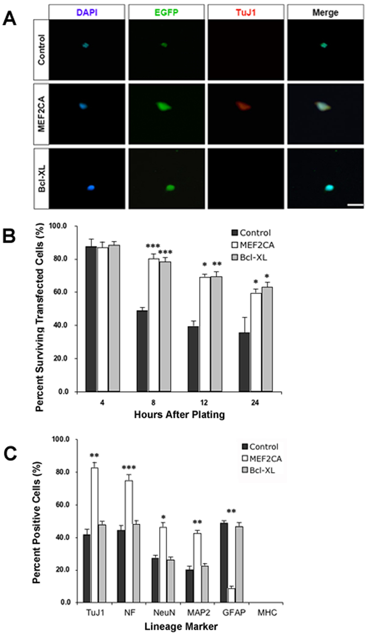
Figure 1. Forced-expression of MEF2CA in ES cells plated at limiting dilution enhances survival and neuronal differentiation.
A, Representative micrographs of murine D3 ES cells transiently transfected with expression plasmids containing EGFP (Control), EGFP/MEF2CA, or EGFP/Bcl-xL under regulation of the nestin promoter. The ESCs were allowed to recover and then plated at a limiting dilution in defined medium without serum or growth factors. Cells were stained with DAPI for nuclear morphology, and TuJ1 as evidence for neuronal differentiation. B, Percentage of surviving cells, treated as described above, quantified after culture for various time periods. C, Percentage of surviving cells 24 h after plating as described above and stained with markers for neurons (TuJ1, NeuN, NF, and MAP-2), astrocytes/progenitor cells (GFAP), or muscle (MHC). Results are expressed as mean + SEM from 3 separate experiments. Statistical significance different from control cells: * p < 0.05, ** p < 0.01, *** p < 0.001. Scale bars, 25 µm.

