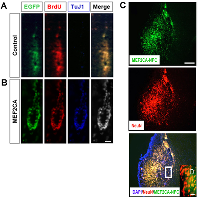
Figure 6. MEF2CA-ESC-derived neuronal progenitor cells injected into mouse stroke model express neuronal markers and migrate into ischemic areas of the brain.
A, B, Four weeks after transplantation, MEF2CA/EGFP-ESC-derived NPCs (B) but not EGFP (control)-expressing progenitors (A) that were located near the injection site stained for the early neuronal marker TuJ1. Cells were co-labeled with BrdU prior to transplantation for identification purposes; scale bar, 25 µm. C, D, By eight weeks after transplantation, many of the injected MEF2CA/EGFP-ESC-derived NPCs had migrated to an area adjacent to the ischemic zone and expressed the mature neuronal marker NeuN (C); scale bar, 200 µm. Colocalization of EGFP and NeuN can be seen more clearly in the high power inset (D); scale bar, 15 µm.

