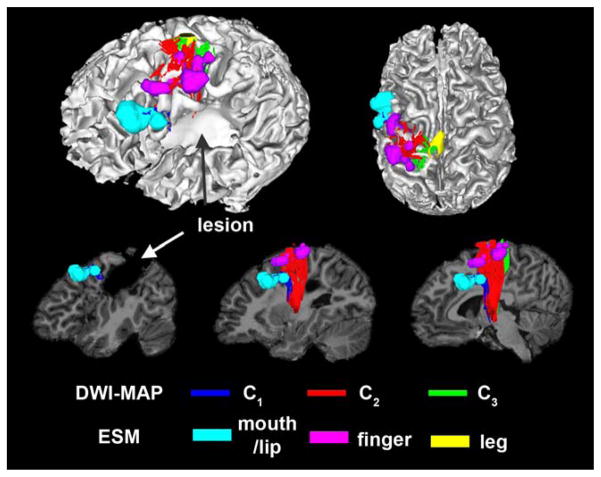
Figure 2.
Automatic detection of three pathways using the DWI-MAP classifier, C1: mouth/lip (blue), C2: finger (red), C3: leg (green) obtained from 9 years old child with focal epilepsy and a lesion involving the pre/post-central gyrus. The ESM sites of mouth/lip (cyan), finger (pink), and leg (yellow) were compared with C1, C2, and C3 of DWI-MPA classifier, respectively. [Color figure can be viewed in the online issue, which is available at wileyonlinelibrary.com.]

