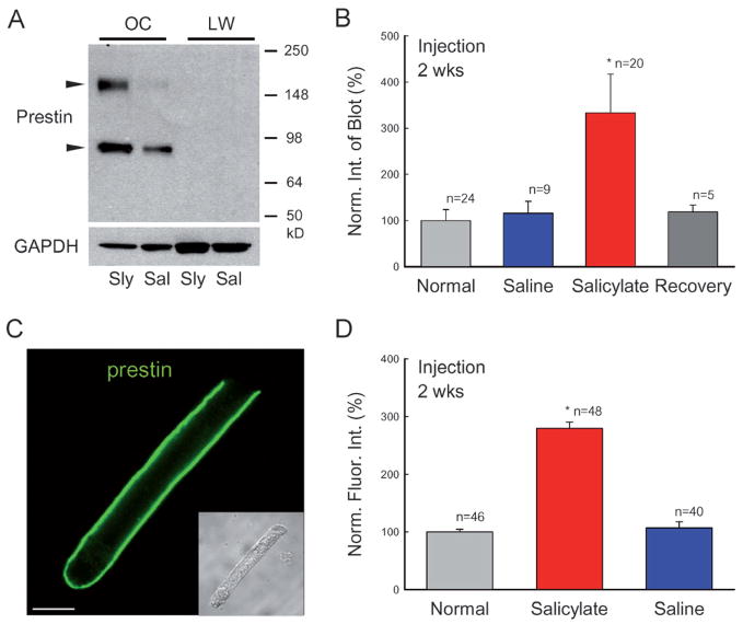
Figure 3.
Increase in prestin protein expression in long-term administration of salicylate. (A) Western blots of the cochlea for prestin. Each well was loaded with whole lysates of one organ of Corti (OC). Cochlear lateral wall tissue (LW) was used as a negative control. Animals were injected with salicylate (Sly) or saline (Sal) for 2 weeks. Prestin immunoreactivity is visible in the organ of Corti samples but is not detected in the cochlear lateral wall samples. Two arrowheads indicate monomer and dimer blots of prestin (86.1 and 172.9 kDa, respectively). (B) Quantitative analysis of prestin Western blots in normal, salicylate and saline groups. The salicylate and saline groups were injected with salicylate or saline for 2 weeks. The recovery group was measured at 4 weeks after stopping salicylate administration (given for 2 weeks). The blot intensity was normalized to the intensity of a normal control. (C) A confocal image of immunofluorescent staining of a guinea pig outer hair cell (OHC) for prestin. (Inset) A Nomarski image. Scale bar: 10 μm. (D) Quantitative analysis of prestin labeling at the OHC lateral wall. The intensity of labeling was normalized to that in the normal control group. Each group contained three guinea pigs; n represents the measured OHC number. Prestin labeling at the OHC lateral wall was increased during long-term administration of salicylate.

