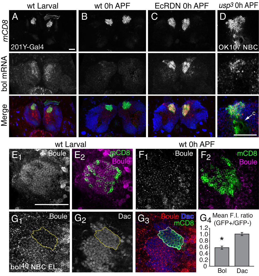
Fig. 4. Boule expression is downregulated by ecdysone signaling in MB neurons at the initiation of metamorphosis.
(A–C) In situ hybridization analysis of Boule mRNA expression in MB γ neurons. Boule mRNA expression in the brain of wt early third instar larvae (A) and 0 hour APF pupae (B). Boule mRNA expression is elevated in EcRDN-expressing MB γ neurons at 0 hours APF (C). Merged images show MB γ neurons in green, boule mRNA in red and DAPI nuclear staining in blue.
(D) Boule mRNA expression in a MB neuroblast clone (NBC) that is homozygous for usp3 at 0 hours APF. Merge shows MB neurons labeled with mCD8::GFP (green) and boule mRNA (red) and DAPI (blue). Arrow denotes calyx region (c), which is devoid of cell bodies.
(E–F) Boule protein expression (magenta) in MB γ neurons (green) in early third instar larvae (E) and 0 hour APF pupae (F), as detected by antibody staining with a rabbit polyclonal antibody against Boule.
(G) Boule protein is decreased in bol40 mutant clones in early third instar larvae. Panels show Boule (G1) and Dachshund (Dac; G2) protein staining, with a merge (G3) showing the MARCM NBC in green, Boule in red and Dac in blue. Yellow dashed line represents the extent of the bol40 clone marked by mCD8::GFP. (G4) Graph of the mean ratio of fluorescence intensity (F.I.) for Boule or Dac protein staining within the bol40 homozygous clones marked by GFP, or in adjacent heterozygous cells (GFP−). Error bars represent s.e.m. The asterisk denotes a p value < 0.002 (two-tailed unpaird T-test, n = 6 MB NBCs). Residual staining with the Boule polyclonal antibody in bol40 clones is likely due to non-specific antibody staining.
Images in (A–D) show confocal z-stacks of 15 µm cryosections of larval or pupal brains showing MB neuron cell bodies marked with mCD8::GFP driven by 201Y-GAL4 or OK107-GAL4. Images for (D & E) are 1 µm optical sections taken from a confocal stack. n ≥ 12 brains for each experiment. Scale bars, 50 µm.

