
Figure 2. DSF-Cu reduces cellular antioxidant capacity to induce ROS and activate redox signaling.
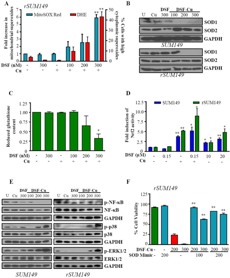
SUM149, rSUM149 cells treated with DSF, DSF+Cu (100-300 nM, 10 μM), Cu alone (10 μM). A, Fold induction of mitochondrial superoxides (white bars) and percentage of cells with high cytoplasmic superoxides (black bars) measured by flow cytometry. B, Immunoblot analysis of SOD1/2. C, Reduced glutathione content relative to untreated (rSUM149 shown). D, Fold induction of Nrf2 activity measured by ARE-responsive luciferase activity. E, Immunoblot analysis of indicated proteins in treated cells at 4 h time point. F, Effect of SOD mimetic (MnTnHexyl-2-Pyp5+, 100-200 μM) on viability measured by trypan blue exclusion assay (rSUM149 shown). *p<0.05, **p<0.005 in all panels. GAPDH and respective total proteins as loading controls.