
Figure 3. DSF induces Cu-dependent apoptosis.
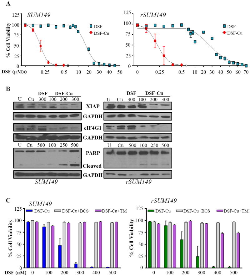
A, Dose-dependent measurement of viability in cells treated with DSF, Cu (10 μM), or DSF-Cu. B, Immunoblot analysis of apoptotic pathway proteins. GAPDH as loading control. C, Viability in the presence of Cu chelators bathocuproine disulphonate (BCS, 100 μM, white bars) or tetrathiomolybdate (TM, 10 μM, gray bars) measured by trypan blue exclusion assay.