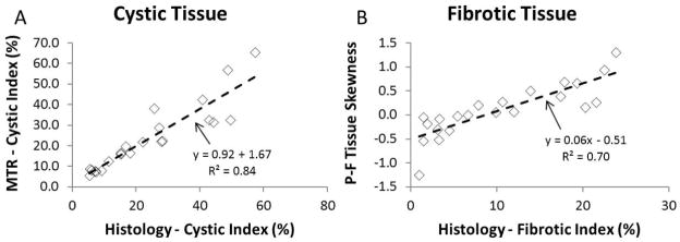
Figure 3.
Panel A: Cystic index computed by GMM approach on MTR image correlated to that measured by histology. Distinguishing the different tissues on MT by the GMM approach had very high accuracy at measuring the degree of cystic burden of each specimen. Panel B: Utilizing a 2-tissue GMM approach, the skewness of the P-F tissue class was computed and correlated with the measured fibrotic index (obtained from the Picro-Sirius Red histology measurement). This correlation was very similar when comparing to the Masson’s trichrome derived fibrotic index.

