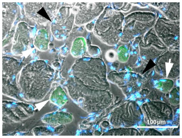
Fig. 7.
Four-year LMN human denervated quadriceps muscle. Immunohistochemical staining with anti-MHCslow shows that both the green-labeled slow type muscle fibers (white arrows) and the fast (larger, not stained muscle fibers noted by black arrowheads) present several central nuclei. Scale bar: 100 μm.

