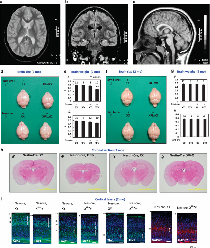
Figure 1. Microcephaly with normal cortical structures in PQBP1 patients and nestin-Cre Pqbp1-cKO mice.
(A–C) Magnetic resonance imaging of a PQBP1-mutated patient showed normal cortical structures with no periventricular heterotopia: (A) horizontal, (B) coronal, (C) sagittal sections.
(D) Macroscopic images of the brain at the age of 2 months. Male nestin-Cre Pqbp1-cKO mouse brains (Nes-Cre; XFloxY) were consistently the smallest among the littermates.
(E) Pqbp1-cKO mice showed reduction of brain weight at 2 months. The bar graph shows the mean + S.E.M for each genotype with the number of mice used indicated above. The mean and S.E.M values are provided in the text. Asterisks indicate significance (p < 0.01) in one way ANOVA with post hoc Bonferroni test.
(F) Macroscopic images of the brain at the age of 2 months. Male synapsin 1-Cre Pqbp1-cKO mouse brains (synapsin1-Cre; XFloxY) were not different from the littermates in size.
(G) The brain weight of the male synapsin-1-Cre Pqbp1-cKO mouse was not different from that of the background control.
(H) Coronal sections of adult brains of nestin-Cre Pqbp1-cKO mouse and littermates (2 months, at −1.82 mm from the bregma in background mice).
(I) Staining for layer-specific markers, Cux1, Foxp1, and Tbr1, together with GAD67, shows preservation of cortical layers in Pqbp1-cKO mice at 2 months.

