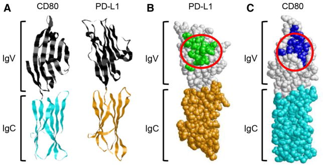
Fig. 2.

CD80, PD-1, PD-L1, and CTLA-4 share common binding sites. a Ribbon structures showing the amino-proximal IgV-like and the amino-distal IgC-like domains of CD80 and PD-L1 (structures are from RCSB Protein Databank (http://www.rcsb.org/pdb/home/home.do); accession numbers 1I8L and 3BIK, respectively). b PD-L1 space-fill structure showing the predicted binding sites for PD-1 (green residues) and CD80 (area circled in red). c CD80 space-fill structure showing the predicted binding sites for CTLA-4 (blue residues) and PD-L1 (area circled in red). Predicted binding sites are based on [17, 48, 49], and identification of amino acid residues that are within 3.9Å of the binding partner
