Fig. 6.
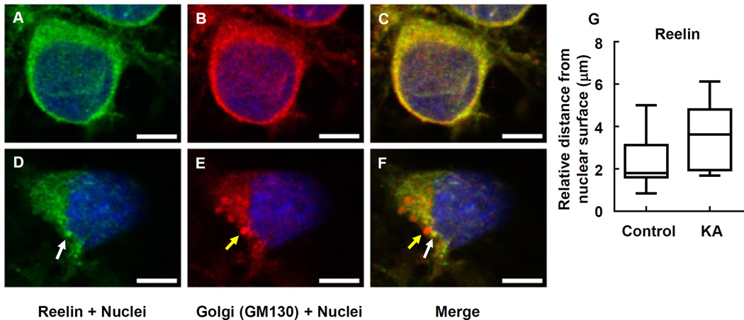
Intracellular Reelin (white arrow) dislocated at Golgi complex (yellow arrow) in response to KA. PRNCs were cultured with KA for 3 days at 37°C. PRNCs were fixed and stained for Reelin and Golgi complex using antibodies against Reelin (green) and GM 130 (red), respectively, and for nuclei using Hoechst 33258 (blue). (A–C) Control, Intracellular Reelin co-localized with Golgi complex, (D–F) PRNCs were incubated with 5 µM KA for 3 days at 37°C. (G) Quantification of Reelin distribution around nuclei corresponded to distance of reconstructed anti-Reelin fluorescent signal from nuclear surface. Scale bars = 5µm.

