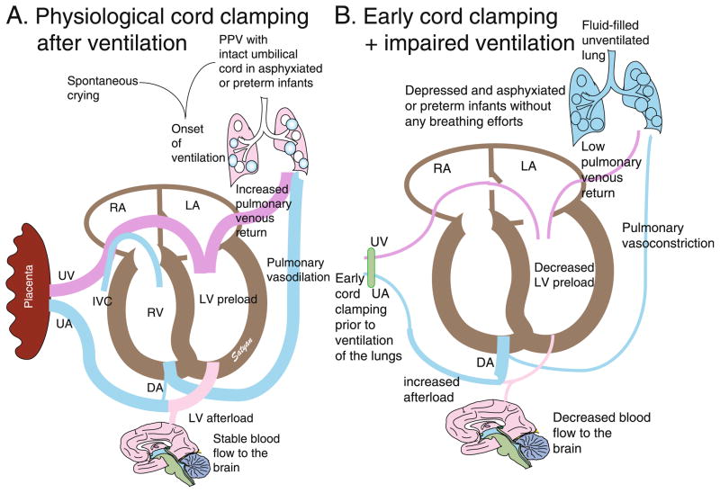
Figure 2. Effects of cord clamping on hemodynamics.
An intact umbilical cord allows continuous umbilical venous flow to the ventricles. With the concomitant initiation of breathing through crying or PPV, PVR decreases allowing increased blood flow to the lungs (decreased right to left shunt through the DA) as well as increased venous return to the LV. The unclamped UA prevents a sudden increase in afterload. This results in improved cardiac output (A). Conversely, immediate cord clamping restricts flow to the ventricles. With failure to establish ventilation, PVR remains high and compromises pulmonary blood flow (increased right to left DA shunt) and venous return to the left ventricle. Thus, decreased filling of the left ventricle (preload) and increased afterload (due to removal of low-resistance placenta) compromise cardiac output (B). DA ductus arteriosus, PPV positive pressure ventilation, LA left atrium, RA right atrium, LV left ventricle, UA umbilical artery, UV umbilical vein. Copyright Satyan Lakshminrusimha.

