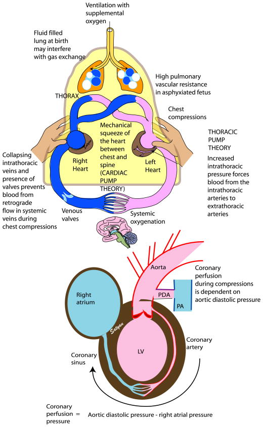
Figure 5. Chest compressions (CC).
During CCs, the extrinsic pressure on the sternum squeezes the heart against the spine leading to antegrade blood flow (cardiac pump theory). During ventilation, generation of increased thoracic pressure results in arterial blood flow from the thorax because intrathoracic pressure exceeds extrathoracic vascular pressure (thoracic pump theory). Flow is restricted to the arterial-to-venous direction because of collapse of veins at the thoracic inlet and venous valves that prevent retrograde flow. Coronary perfusion pressure (CPP) is a key determinant in return of spontaneous resuscitation and is dependent on aortic diastolic and right atrial pressure. In the presence of a PDA, CCs may be less effective as aortic diastolic pressure may be decreased with blood shunting from the aorta into the pulmonary artery, thus decreasing CPP. LV left ventricle, PA pulmonary artery, PDA patent ductus arteriosus. Copyright Satyan Lakshminrusimha.

