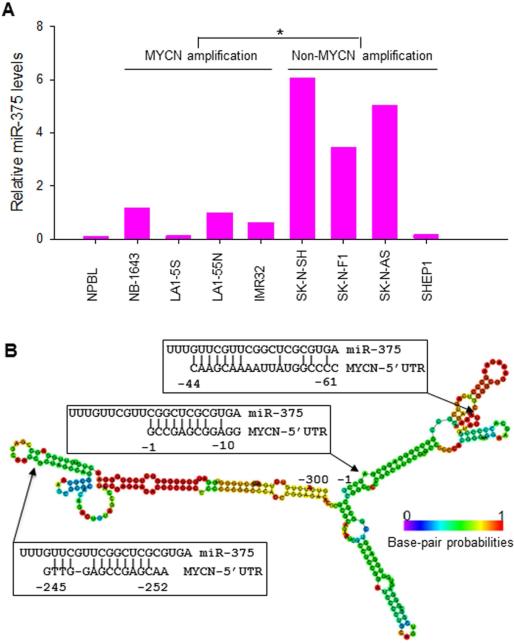
Figure 1.
A, expression of miR-375 in 8 NB cell lines with or without MYCN amplification and in normal peripheral blood lymphocyte (NPBL), as detected by qRT-PCR. Results are given as average levels of three measurements, normalized to the internal control RUN24. The mean levels of MYCN amplified and non-MYCN-amplified groups were compared with a significant difference, *p<0.01. B, the putative miR-375 binding sites in the MYCN 5’-UTR (−1 to −300). Arrows indicate the MYCN 5’-UTR sequences that are complementary to miR-375. The colors of the secondary structure of the MYCN 5’-UTR indicated the propensity of the individual nucleotides to participate in base pairs. The scale ranges from red (highest probability) to blue-violet (lowest probability).

