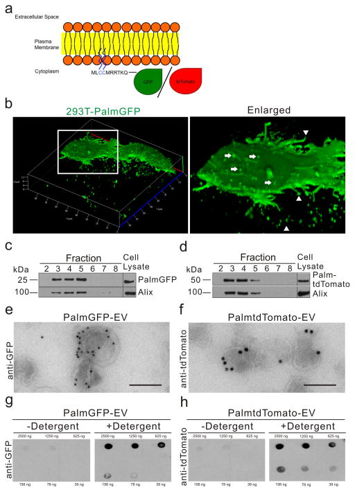
Figure 1. Palmitoylated-GFP and -tdTomato are trafficked to the plasma membrane and EVs.
(a) Schematic diagram of EV membrane labeling with palmitoylated GFP (PalmGFP) or tdTomato (PalmtdTomato). (b) Live-cell confocal microscopy of stable PalmGFP-expressing 293T cells releasing EVs. Left panel: 3D reconstruction of confocal Z-stack images demonstrating EV release from stable 293T-PalmGFP cells into surrounding regions. Right panel: Enlarged image of boxed region, showing budlike structure from the surface (arrows) as well as processes extending from cells (arrow heads). (c, d) Western blot analysis of protein extracted from fractions following sucrose gradient centrifugation of isolated EVs. PalmGFP (c) and PalmtdTomato (d) were detected in fractions exhibiting the EV marker, Alix (95 kDa). (e, f) Transmission electron micrograph (TEM) showing PalmGFP and PalmtdTomato labeling of EVs on the membrane following immunolabeling with anti-GFP (e) or anti-tdTomato (f) and secondary gold-conjugated secondary antibodies. Bar, 100 nm. (g) Demonstration that PalmGFP and PalmtdTomato labels the inner membrane of EVs. PalmGFP- or PalmtdTomato EVs were dot blotted on nitrocellulose membrane in a dose range followed by immunoprobing with anti-GFP or anti-tdTomato, respectively, and horseradish peroxidase (HRP)-conjugated secondary antibodies in the presence or absence of detergent [0.1% (v/v) Tween20] for chemiluminescence detection.

