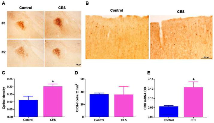
Fig. 4.
Corticotropin releasing hormone (CRH) expression is augmented in the amygdala of CES rats. (A) Representative photomicrographs of amygdalae after immunohistochemistry using an antiserum directed against CRH. CRH immunoreactivity (ir) was enhanced in the amygdala of CES rats compared with controls at P45 as shown in C: Semi-quantitative analysis of CRH expression levels revealed a significant increase of the optical density of CRH-ir in rats that experienced CES, (p = 0.048). (B) In frontoparietal-cortex, CRH-ir cell number and density did not differ significantly in the same control and CES rats distinguished by amygdala expression. Representative micrographs show the typical distribution of CRH-expressing cortical interneurons. (D) A graph depicting the number of cells expressing CRH above detection levels in an area of 2 square mm (controls: 36.3 ± 2.1; CES: 35.8 ± 13.2; p = 0.97, t-test with Welch correction for unequal variance). Cell numbers were not used in amygdala sections because, as apparent in the photos in A, most CRH in this nucleus is found in fibers. (E) At the age of onset of spasm-like events (pre-weaning or infancy, P19), CRH mRNA expression levels were borderline higher in CES rats compared to controls (CES: 0.12 ± 0.01; controls: 0.08 ± 0.002, n= 3–4 per group; p =0.04, Mann-Whitney test. Scale: 250 μm.

