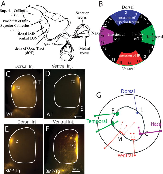
Figure 1.
Projections from ventral, but not dorsal retina are inappropriate in BMP transgenic mice at P8. A. Schematic of mouse visual system, showing the major retinal projections, including the dorsal and ventral LGN and the SC. The accessory optic system is not shown. The ‘delta of the Optic Tract’ is the area of the ascending optic tract before it enters the ventral LGN. The ‘brachium of the Superior Colliculus’ is the area of the optic tract between the LGN and SC, just before the ganglion cell axons enter the SC. B. The retina is assigned coordinates based on the insertion of the four major occulomotor muscles, namely the superior, inferior, lateral, and medial recti (SR, IR, LR and MR), see Plas et al., 2005. C. A focal injection into dorsal retina of a WT mouse labels a target zone (TZ) in lateral SC. D. A focal injection into ventral retina of a WT mouse labels a TZ in medial SC. E. In BMP transgenic mice, the projection from dorsal retina is similar to that in WT mice and topographically correct. F. Projections from ventral retina in BMP transgenic mice show ‘ectopic spots’ (arrows) lateral to the expected TZ. G. Summary cartoon depicting graphically the frequency of inappropriate projections to the SC as a function of the position of the retinal dye injection in the BMP transgenic mouse. Dorsal retinal injections lead to normal collicular projections to lateral (L) colliculus (blue). Ventral retinal injections lead to a medial target zone (M) in colliculus and many ectopic spots (red). Temporal (green) and nasal (purple) retinal injections are largely normal, with some cases of inappropriate projections to the colliculus.

